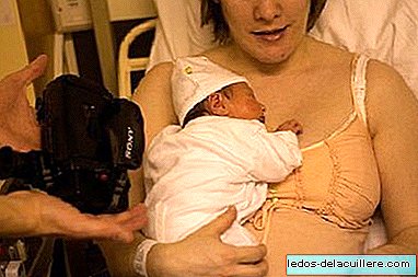
Retrolenticular or retrocristalinian fibroplasia or Retinopathy of prematurity is an abnormal development of blood vessels in the retina of the eye In a premature baby. Some cases of this eye disease are mild and correct themselves, but others require surgery to prevent vision loss or blindness.
How this retinal disorder occurs, what are the risk factors, treatment and its consequences we will talk about below.
Retinopathy causes the growth of abnormal blood vessels in the retina, the layer of nerve tissue in the eye that allows vision. This growth can cause the retina to detach from the back of the eye in the most severe cases.
The blood vessels of the retina begin to develop three months after conception and complete their development at the time of birth after 38 weeks. If a baby is born too early, eye development is not complete., it is altered and a multiplicity of factors can first cause an arrest and then an abnormal growth of the retinal vessels.
The result is that the fragile vessels may have losses, causing bleeding in the eye. Likewise, scar tissue can develop and detach the retina from the inner surface of the eye, even in the most severe cases causing vision loss.
This pathology usually occurs in two phases (which overlap to some degree):
An acute phase, in which normal vasculogenesis is interrupted and retinal response to an injury is observed.
A phase of late or chronic proliferation of membranes towards the vitreous, during which there are tractional detachments of the retina, ectopia and scarring of the macula, which leads to a considerable loss of vision, and can lead to total loss.

Risk factors and treatment of retinopathy of prematurity
In addition to prematurity and low birth weight (less than 1750 grams are considered), others risk factor's they can be apnea or brief respiratory arrest, heart disease, a high level of carbon dioxide (CO2) in the blood, infection, low level of oxygen or acidity in the blood, shortness of breath, low heart rate (bradycardia), transfusions .
Currently, new care to treat premature babies means that the risk of developing retinopathy depends on the degree of prematurity, being the most affected those more premature babies (especially less than 30 weeks of gestation), small and sick .
The incidence of retinopathy of prematurity (ROP) is inversely proportional to birth weight, estimated to occur in 30% of children under 1500 grams. It is the case that with the current advances in neonatology the survival of these babies has increased of 1500 grams or less, which are the children who have the highest risk of suffering retinopathy of prematurity.
Babies born prematurely have a retinal exam after four weeks (later if they are large premature) to see how the eye vessels are developing and to act accordingly.
However, there are those who point out that Checks should be made even before four weeks, since early treatment has shown that it improves the chances that the baby has normal vision and cases of severe myopia or blindness are avoided.
Treatment may include cryotherapy (freezing) and laser therapy (photocoagulation) can be used to prevent complications of retinopathy of advanced prematurity. Surgery is necessary if the retina detaches.
Among the decalogue of rights of premature babies is the prevention of blindness due to retinopathy of prematurity: a premature child needs ophthalmological controls from the first weeks of life.
In addition, as premature babies have the possibility of presenting other ocular alterations (myopia, strabismus, delayed visual maturation ...), further ophthalmologic examinations must be carried out.
This way you can achieve that most premature babies with retinopathy recover without problems durable visuals. However, the most severe cases are approximately 1 in 10 babies who will develop a more severe retinal disease resulting in severe myopia and blindness.